Ultrasound Transducer
The active component needed to make ultrasound images is the ultrasound transducer. The origins of which come from experiments performed by Pierre and Jacques Curie in 1880, France. Working with quartz crystals, they found that pressure on them created an electric pulse of energy. Called the Piezoelectric effect, it was the reverse of a known phenomenon where an electric pulse changed the properties of a material. See Pierre Curie Human hearing is from about 20 cycles per second or 20HZ (a low hum), Below that is infrasonic, to about 20,000 cycles per second or 20KHZ. Above that is ultrasonic.

The transducer is a hand held device that is placed on the skin over the subject being observed. A gel is applied on the skin because ultrasound works best through liquid. It transmits an alternating pulse of energy into the tissue and the sound is absorbed, reflected, or scattered. Between pulses, is receives the echos and sends the information back to the computer. The computer then translates the information by subtracting the pulse echoed back from the known level of energy transmitted.

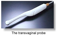
There are different types of transducers for different purposes. Some transmit waves in a wide band to show a large object, such as an organ, or fetus. Some have a different array for cancer, cysts, fibroids, fluid blockages etc. Then there are the types designed for internal exams, such as for
Transvaginal ultrasound
or
prostate ultrasound
Some have a very narrow pulse to show between the ribs to inspect the heart. There are many specialized types of
ultrasound transducers. back to what is ultrasound

Genesis ultrasound machine Home Page


