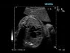
Heart Ultrasound
Heart ultrasound is known as an echocardiogram, which is a sonogram of the heart and can be called cardiac ultrasound or cardiology ultrasound. We can use 2D, which takes images that are like slices of a loaf of bread. This can show abnormalities in a very distinct manner. We do
3D,
which shows pictures of the heart like a sculpture, and
4D Doppler,
which makes images in real time movement so we can see blood flows and obstructions.

This allows assessment of cardiac valves and their function or malfunction, as the case may be. We can view leakage between valves and chambers called valvular regurgitation, as well as overall output. There are many variations of this procedure, including a cardiac perfusion scan. A heart ultrasound measures the amount of blood in a heart muscle at rest and during exercise. This is commonly done to find out what is causing chest pain. There are numerous conditions and abnormalities detected by an exam.
In August, 2008 Austria, a study was done by researchers by doing repeated ultrasound scans of the carotid artery
See Carotid ultrasound
to determine heart attack and stroke risk from increased plaque build up. The carotid arteries are in the neck. They studied cholesterol, fat and other materials found in arteries. Patients with unstable amounts of plaque were more likely to have medical complications. The instability is caused by factors such as diet and exercise. 600,000 have their first stroke and another 600,000 have their first heart attack each year. But "the determination of the degree of stenosis alone is insufficient to predict patients' risk," says Markus Reiter, MD, a physician at Medical University of Vienna, and the study's lead author.
They studied patients who had no symptoms, but had high risk factors. these people had multiple risk factors, such as smoking, diabetes, high blood pressure, high cholesterol, or known blockages in other blood vessels such as the coronary arteries. The focus was on 574 patients with plague build up. Multiple heart ultrasound scans were done over a period of six to nine months.
In follow up studies done after three years, the GSM levels decreased in 230 patients, or 40%, and increased in 344, or 60%. Reiter used ultrasound images and a computer-assisted evaluation (called gray-scale median or GSM) to evaluate the darkness of the plaque and its density. If plaque appears darker, it has a low GSM and is thought to be unstable. Those in the lowest GSM group, with the darkest plaque, were about 1.7 times more likely to have a heart attack or stroke than those whose GSM went up the most, reflecting less dense plaque.
It is generally agreed that more study is needed, in that this one was done with only patients with high risk factors and not from the general population. However, It does seem to indicate that people need to watch what they eat and get regular exercise.
For more, you can click to
heart ultrasound at Web MD.
Back to types of ultrasound
Genesis ultrasound machine Home Page
Thanks to:
Markus Reiter, MD, a physician at Medical University of Vienna
By Kathleen Doheny WebMD Health News
Reviewed by Elizabeth Klodas, MD, FACC
Cleveland Clinic


